
Dermatophytosis includes several distinct clinical entities, depending on the anatomic site and etiologic agents involved. Clinically, the conditions include tinea capitis, tinea favosa (favus resulting from infection by Trichophyton schoenleinii), tinea corporis (ringworm of glabrous skin), tinea imbricata (ringworm resulting from infection by Trichophyton concentricum), tinea cruris (ringworm of the groin), tinea unguium or onychomycosis (ringworm of the nail), tinea pedis (ringworm of the feet), tinea barbae (ringworm of the beard), and tinea manuum (ringworm of the hand).
Pathophysiology
Tinea capitis is caused by fungi of species of genera Trichophyton and Microsporum.
Tinea capitis is the most common pediatric dermatophyte infection worldwide. The age predilection is believed to result from the presence of Pityrosporum orbiculare (Pityrosporum ovale), which is part of normal flora, and from the fungistatic properties of fatty acids of short and medium chains in postpubertal sebum.
Causative agents of tinea capitis include keratinophilic fungi termed dermatophytes. These molds usually are present in nonliving cornified layers of skin and its appendages and sometimes are capable of invading the outermost layer of skin, stratum corneum, or other keratinized skin appendages derived from epidermis, such as hair and nails.
Dermatophytes are among the most common infectious agents of humans, causing a variety of clinical conditions that are collectively termed dermatophytosis. From the site of inoculation, the fungal hyphae grow centrifugally in the stratum corneum. The fungus continues downward growth into the hair, invading keratin as it is formed. The zone of involvement extends upwards at the rate at which hair grows, and it is visible above the skin surface by days 12-14. Infected hairs are brittle, and by the third week, broken hairs are evident.
Three types of in vivo hair invasion are recognized.
- Ectothrix invasion is characterized by the development of arthroconidia on the exterior of the hair shaft. The cuticle of the hair is destroyed, and infected hairs usually fluoresce a bright greenish-yellow color under a Wood lamp ultraviolet light. Common agents include Microsporum canis, Microsporum gypseum, Trichophyton equinum, and Trichophyton verrucosum.
- Endothrix hair invasion is characterized by the development of arthroconidia within the hair shaft only. The cuticle of the hair remains intact and infected hairs do not fluoresce under a Wood lamp ultraviolet light. All endothrix-producing agents are anthropophilic (eg, Trichophyton tonsurans, Trichophyton violaceum).[1]
- Favus, usually caused by T schoenleinii, produces favuslike crusts or scutula and corresponding hair loss.
Epidemiology
Frequency
United States
Occurrence of the disease is no longer registered by public health agencies; therefore, true incidence is unknown. The reported peak incidence occurs in school-aged African American male children.Tinea capitis is predominantly a disease of preadolescent children. It accounts for up to 92.5% of dermatophytoses in children younger than 10 years. The disease is rare in adults, although occasionally, it may be found in elderly patients. Tinea capitis occurrence is widespread in some urban areas in the United States.
International
Tinea capitis is widespread in some urban areas, particularly in children of Afro-Caribbean extraction, in North America, Central America, and South America. It is common in parts of Africa and India.[2, 3, 4, 5] In Southeast Asia, the rate of infection has been reported to have decreased dramatically from 14% (average of male and female children) to 1.2% in the last 50 years because of improved general sanitary conditions and personal hygiene. In northern Europe, the disease is sporadic.In the United Kingdom and North America, T tonsurans accounts for greater than 90% of cases of infection .[6] In the nonurban communities, sporadic infections acquired from puppies and kittens are due to M canis, which accounts for less than 10% of cases in the United Kingdom. Occasional infection from other animal hosts (eg, T verrucosum from cattle) occurs in rural areas.
Mortality/Morbidity
Classification and severity of tinea capitis depend on the site of formation of their arthroconidia.- Ectothrix infection is defined as fragmentation of the mycelium into conidia around the hair shaft or just beneath the cuticle of the hair, with destruction of the cuticle. Inflammatory tinea related to exposure to a kitten or puppy usually is a fluorescent small spore ectothrix. Some mild ringworm or prepubertal tinea capitis infections are of the ectothrix type, also termed the gray-patch type (microsporosis; see the image below). Some ectothrix infections involute during the normal course of disease without treatment. Depending on the extent of associated inflammation, lesions may heal with scarring.
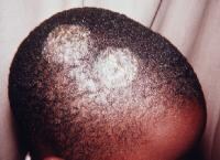 Gray-patch
ringworm (microsporosis) is an ectothrix infection or prepubertal tinea
capitis seen here in an African American male child. Gray patch refers
to the scaling with lack of inflammation, as noted in this patient.
Hairs in the involved areas assume a characteristic dull, grayish,
discolored appearance. Infected hairs are broken and shorter. Papular
lesions around hair shafts spread and form typical patches of ring
forms, as shown. Culture from the lesional hair grew Microsporum canis.
Gray-patch
ringworm (microsporosis) is an ectothrix infection or prepubertal tinea
capitis seen here in an African American male child. Gray patch refers
to the scaling with lack of inflammation, as noted in this patient.
Hairs in the involved areas assume a characteristic dull, grayish,
discolored appearance. Infected hairs are broken and shorter. Papular
lesions around hair shafts spread and form typical patches of ring
forms, as shown. Culture from the lesional hair grew Microsporum canis. - Endothrix infections are noted in which arthrospores are present within the hair shaft in both anagen and telogen phases, contributing to the chronicity of the infections. Endothrix infections tend to progress, become chronic, and may last into adult life. Lesions can be eradicated by systemic antifungal treatment. Since the organisms usually remain superficial, little potential for mortality exists. Disseminated systemic disease has been reported in patients who are severely immunocompromised.
Sex
The incidence of tinea capitis may vary by sex, depending on the causative fungal organism. Microsporum audouinii –related tinea capitis has been reported to be up to 5 times more common in boys than in girls. After puberty, however, the reverse is true, possibly because of women having greater exposure to infected children and possibly because of hormonal factors. In infection by M canis, the ratio varies, but the infection rate usually is higher in boys. Girls and boys are affected equally by Trichophyton infections of the scalp, but in adults, women are infected more frequently than are men.Age
Tinea capitis occurs primarily in children and occasionally in other age groups. It is seen most commonly in children younger than 10 years. Peak age range is in patients aged 3-7 years.


4 comments:
Nice work you are doing here brother. I'm a good fan of your work.
Well done here. See Also ----> Road To Success And Wellness.
I'm 15 years old. I was born with HIV my mother passed away because of the HIV infection And I regret why i never met Dr Itua he could have cured my mum for me because as a single mother it was very hard for my mother I came across Dr itua healing words online about how he cure different disease in different races diseases like HIV/Aids Herpes,Parkison,Copd,Epilepsy,Shingles,Cold Sore,Infertility, Chronic Fatigues Syndrome,Fibromyalgia, Diabetes Hepatitis even Cancer I was so excited but frighten at same time because I haven't come across such thing article online then I contacted Dr Itua on Mail drituaherbalcenter@gmail.com I also chat with him on what's app +2348149277967 he tells me how it works then I tell him I want to proceed I paid him so swiftly Colorado post office I receive my herbal medicine within 4/5 working days he gave me guild lines to follow and here am I living healthy again can imagine how god use men to manifest his works am I writing in all articles online to spread the god work of Dr Itua Herbal Medicine,He's a Great Man.
HOW I GOT CURED OF HERPES VIRUS.
Hello everyone out there, i am here to give my testimony about a herbalist called dr imoloa. i was infected with herpes simplex virus 2 in 2013, i went to many hospitals for cure but there was no solution, so i was thinking on how i can get a solution out so that my body can be okay. one day i was in the pool side browsing and thinking of where i can get a solution. i go through many website were i saw so many testimonies about dr imoloa on how he cured them. i did not believe but i decided to give him a try, i contacted him and he prepared the herpes for me which i recieved through DHL courier service. i took it for two weeks after then he instructed me to go for check up, after the test i was confirmed herpes negative. am so free and happy. so, if you have problem or you are infected with any disease kindly contact him on email--- drimolaherbalmademedicine@gmail.com. or / whatssapp --+2347081986098.
This testimony serve as an expression of my gratitude. he also have herbal cure for COLD SORE, SHINGLES, CANCER, HEPATITICS A, B. DIABETES 1/2, HIV/AIDS, CHRONIC PANCERATIC, CHLAMYDIA, ZIKA VIRUS, EMPHYSEMA, LOW SPERM COUNT, ENZYMA, COUGH, ULCER, ARTHRITIS, LEUKEMIA, LYME DISEASE, ASTHMA, IMPOTENCE, BARENESS/INFERTILITY, WEAK ERECTION, PENIS ENLARGEMENT. AND SO ON.
Post a Comment